

What Are Two Types Of Body Symmetry In Animals What Are Germ Layers
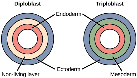
Nearly animal species undergo a separation of tissues into germ layers during embryonic development. Recall that these germ layers are formed during gastrulation, and that they are predetermined to develop into the animal's specialized tissues and organs. Animals develop either 2 or iii embryonic germs layers (Figure). The animals that brandish radial symmetry develop two germ layers, an inner layer (endoderm) and an outer layer (ectoderm). These animals are called diploblasts. Diploblasts accept a non-living layer between the endoderm and ectoderm. More circuitous animals (those with bilateral symmetry) develop three tissue layers: an inner layer (endoderm), an outer layer (ectoderm), and a middle layer (mesoderm). Animals with three tissue layers are called triploblasts.
Fine art Connexion
Which of the following statements about diploblasts and triploblasts is false?
- Animals that display radial symmetry are diploblasts.
- Animals that display bilateral symmetry are triploblasts.
- The endoderm gives ascent to the lining of the digestive tract and the respiratory tract.
- The mesoderm gives rise to the central nervous organization.
Each of the iii germ layers is programmed to give ascent to item trunk tissues and organs. The endoderm gives rise to the lining of the digestive tract (including the stomach, intestines, liver, and pancreas), too as to the lining of the trachea, bronchi, and lungs of the respiratory tract, along with a few other structures. The ectoderm develops into the outer epithelial covering of the torso surface, the central nervous system, and a few other structures. The mesoderm is the third germ layer; information technology forms betwixt the endoderm and ectoderm in triploblasts. This germ layer gives rise to all muscle tissues (including the cardiac tissues and muscles of the intestines), connective tissues such equally the skeleton and blood cells, and most other visceral organs such as the kidneys and the spleen.
Presence or Absence of a Coelom
Further subdivision of animals with three germ layers (triploblasts) results in the separation of animals that may develop an internal trunk cavity derived from mesoderm, chosen a coelom, and those that practice not. This epithelial cell-lined coelomic cavity represents a space, usually filled with fluid, which lies between the visceral organs and the body wall. It houses many organs such as the digestive system, kidneys, reproductive organs, and middle, and contains the circulatory organization. In some animals, such as mammals, the part of the coelom chosen the pleural crenel provides space for the lungs to expand during breathing. The evolution of the coelom is associated with many functional advantages. Primarily, the coelom provides cushioning and shock absorption for the major organ systems. Organs housed within the coelom can grow and move freely, which promotes optimal organ development and placement. The coelom too provides infinite for the diffusion of gases and nutrients, equally well as body flexibility, promoting improved creature move.
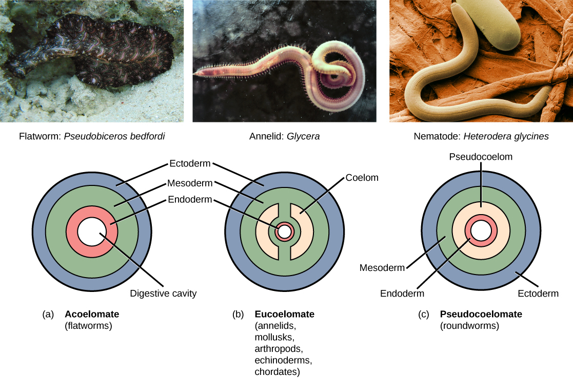
Triploblasts that practice not develop a coelom are called acoelomates, and their mesoderm region is completely filled with tissue, although they do still have a gut crenel. Examples of acoelomates include animals in the phylum Platyhelminthes, also known as flatworms. Animals with a true coelom are called eucoelomates (or coelomates) (Figure). A true coelom arises entirely within the mesoderm germ layer and is lined by an epithelial membrane. This membrane as well lines the organs inside the coelom, connecting and holding them in position while allowing them some gratuitous motion. Annelids, mollusks, arthropods, echinoderms, and chordates are all eucoelomates. A third group of triploblasts has a slightly different coelom derived partly from mesoderm and partly from endoderm, which is establish between the 2 layers. Although nonetheless functional, these are considered false coeloms, and those animals are chosen pseudocoelomates. The phylum Nematoda (roundworms) is an example of a pseudocoelomate. True coelomates can be further characterized based on sure features of their early embryological development.
Embryonic Development of the Mouth
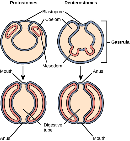
Bilaterally symmetrical, tribloblastic eucoelomates can exist farther divided into two groups based on differences in their early embryonic development. Protostomes include arthropods, mollusks, and annelids. Deuterostomes include more circuitous animals such equally chordates only also some simple animals such as echinoderms. These two groups are separated based on which opening of the digestive crenel develops start: oral fissure or anus. The word protostome comes from the Greek word meaning "mouth start," and deuterostome originates from the word significant "mouth 2nd" (in this case, the anus develops showtime). The mouth or anus develops from a structure called the blastopore (Figure). The blastopore is the indentation formed during the initial stages of gastrulation. In later stages, a second opening forms, and these two openings will somewhen give rise to the mouth and anus (Effigy). It has long been believed that the blastopore develops into the oral cavity of protostomes, with the second opening developing into the anus; the opposite is truthful for deuterostomes. Recent testify has challenged this view of the development of the blastopore of protostomes, however, and the theory remains under debate.
Another stardom between protostomes and deuterostomes is the method of coelom formation, beginning from the gastrula stage. The coelom of about protostomes is formed through a process called schizocoely, meaning that during development, a solid mass of the mesoderm splits apart and forms the hollow opening of the coelom. Deuterostomes differ in that their coelom forms through a procedure chosen enterocoely. Hither, the mesoderm develops as pouches that are pinched off from the endoderm tissue. These pouches eventually fuse to class the mesoderm, which then gives rise to the coelom.
The earliest distinction betwixt protostomes and deuterostomes is the type of cleavage undergone by the zygote. Protostomes undergo screw cleavage, meaning that the cells of ane pole of the embryo are rotated, and thus misaligned, with respect to the cells of the opposite pole. This is due to the oblique angle of the cleavage. Deuterostomes undergo radial cleavage, where the cleavage axes are either parallel or perpendicular to the polar axis, resulting in the alignment of the cells between the two poles.
There is a 2nd distinction between the types of cleavage in protostomes and deuterostomes. In addition to spiral cleavage, protostomes also undergo determinate cleavage. This means that even at this early stage, the developmental fate of each embryonic cell is already determined. A cell does not take the power to develop into whatever cell blazon. In contrast, deuterostomes undergo indeterminate cleavage, in which cells are non yet pre-determined at this early stage to develop into specific jail cell types. These cells are referred to as undifferentiated cells. This feature of deuterostomes is reflected in the existence of familiar embryonic stem cells, which take the power to develop into any jail cell type until their fate is programmed at a later on developmental stage.
Evolution Connection
The Development of the CoelomOne of the first steps in the nomenclature of animals is to examine the animal's body. Studying the body parts tells us non just the roles of the organs in question but also how the species may accept evolved. One such structure that is used in classification of animals is the coelom. A coelom is a body cavity that forms during early on embryonic development. The coelom allows for compartmentalization of the body parts, then that unlike organ systems can evolve and nutrient transport is possible. Additionally, because the coelom is a fluid-filled cavity, information technology protects the organs from shock and pinch. Simple animals, such every bit worms and jellyfish, practise non have a coelom. All vertebrates have a coelom that helped them evolve circuitous organ systems.
Animals that do not take a coelom are chosen acoelomates. Flatworms and tapeworms are examples of acoelomates. They rely on passive improvidence for nutrient transport beyond their body. Additionally, the internal organs of acoelomates are not protected from burdensome.
Animals that have a true coelom are called eucoelomates; all vertebrates are eucoelomates. The coelom evolves from the mesoderm during embryogenesis. The abdominal cavity contains the breadbasket, liver, gall bladder, and other digestive organs. Another category of invertebrates animals based on body crenel is pseudocoelomates. These animals take a pseudo-crenel that is not completely lined past mesoderm. Examples include nematode parasites and pocket-size worms. These animals are thought to have evolved from coelomates and may have lost their ability to grade a coelom through genetic mutations. Thus, this pace in early on embryogenesis—the formation of the coelom—has had a big evolutionary impact on the various species of the animal kingdom.
Source: https://oertx.highered.texas.gov/courseware/lesson/1747/student/?task=3
Posted by: mccabethiss1969.blogspot.com
0 Response to "What Are Two Types Of Body Symmetry In Animals What Are Germ Layers"
Post a Comment